Description
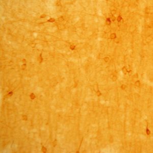
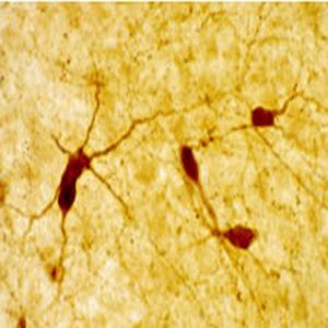
The antiserum was quality control tested using standard immunohistochemical methods. The antiserum demonstrates strongly positive labeling of rat thalamus, cortex, and hippocampus.
The antiserum has been characterized as specific to parvalbumin; please see reference listed below. Recommended primary dilution is 1/5,000 – 1/8,000 in PBS/0.3% Triton X-100 – biotin/avidin-HRP Technique.
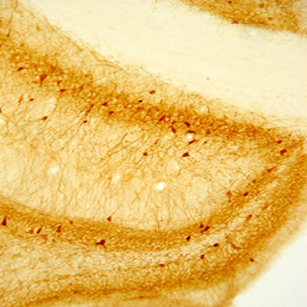
Photo Description: IHC image of the rat hippocampus staining for parvalbumin. The tissue was fixed with 4% formaldehyde in phosphate buffer, before being removed and prepared for vibratome sectioning. Floating sections were incubated at RT in 10% goat serum in PBS, before standard IHC procedure. Primary antibody was incubated at 1:5000 for 48 hours, goat anti-guinea pig secondary was subsequently added for 1 hour after washing with PBS. Light microscopy staining was achieved with standard biotin-streptavidin/HRP procedure and DAB chromogen.
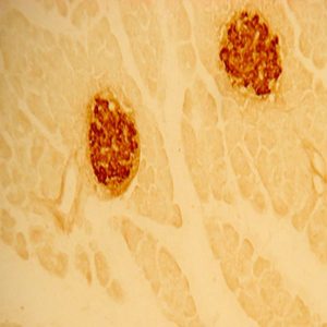
The second image above entitled “D.” is parvalbumin (RED) interneurons pictured in mouse striatum, GREEN is GFP expressed by medium spiny neurons, provided Alipi Naydenov, Neurobiology and Behavior, Stella Lab University of Washington (see review below).
Host: Guinea Pig
Quantity / Volume: 100 µL
State: Lyophilized Whole Serum
Reacts With: Rat
Availability: In Stock
Alternate Names: Parvalbumin alpha; Pva; Parvalbumin (calcium binding protein); PALB1, PC255L (Millipore Cat #), anti-parvalbumin
Gene Symbol: Pvalb
RRID: AB_572259
Database Links:
Entrez Gene: 5816 Human
Entrez Gene: 697272 Monkey
Entrez Gene: 19293 Mouse
Entrez Gene: 102094134 Pigeon
Entrez Gene: 25269 Rat
Technical Sheets
This product contains the preservative sodium azide. The concentration percent of the sodium azide is ≤ .09%. Although this hazardous substance is a concentration below that required for the preparation of a Material Safety Data Sheet, we created a standard MSDS for your records.
Download Data SheetDownload MSDS
Reviews
Want to leave a review? Please click here to send us your review.
Antibody Performed Very Well
The second image above labeled “D.” is parvalbumin (RED) interneurons pictured in mouse striatum, GREEN is GFP expressed by medium spiny neurons. Parvalbumin was used at 1:1000. This antibody performed very well. Scale bar = 25 microns.
Immunohistochemistry: Mice were euthanized and perfused with PFA (4% in PBS), post fixed for 24 hrs, and their brains cryoprotected in 15% sucrose (24 hrs) followed by 30% sucrose (48 hrs). Coronal sections that included the corticostriatal, globus pallidus, or substantia nigra regions (30 um) were prepared using a microtome and then stored in PBS at 4C until processing. Sections were processed/stained in parallel as follows: Free floating sections were rinsed 3x with PBS and incubated 90 min at room temperature (RT) in PBS supplemented with donkey serum (5%) and Triton X-100 (1%). Primary antibody combinations were prepared as a master stock in PBS supplemented with donkey serum (2.5%) and Triton X-100 (0.5%) and applied to sections for 72 hrs at 4oC with gentle agitation. Sections were then rinsed 8x with PBS supplemented with Tween-20 (0.05%, at RT) and incubated with a secondary antibodies diluted in PBS supplemented with donkey serum (2.5%) and Triton X-100 (0.5%) for 1 hr at RT with gentle agitation, followed by 7 rinses with PBS and one rinse with deionized water. Sections were mounted onto slides and allowed to dry for ~18 hrs, after which cover slips were mounted with Vectashield and sealed with nail polish.
Review submitted by:
Alipi Naydenov
Neurobiology and Behavior, Stella Lab
University of Washington