Description
The ImmunoStar Oxytocin antiserum was quality control tested using standard immunohistochemical methods. The antiserum demonstrates strongly positive labeling of rat hypothalamus using indirect immunofluorescent and biotin/avidin-HRP techniques. Recommended primary dilution is 1/4,000 – 1/8,000 in PBS/0.3% Triton X-100 – Bn/Av-HRP.
Staining is completely eliminated by pretreatment of 1 mL of the diluted antibody with 5 µg of Oxytocin. Pretreatment of 1 mL of the diluted antibody with as much as 100 µg of vasopressin does not diminish staining.
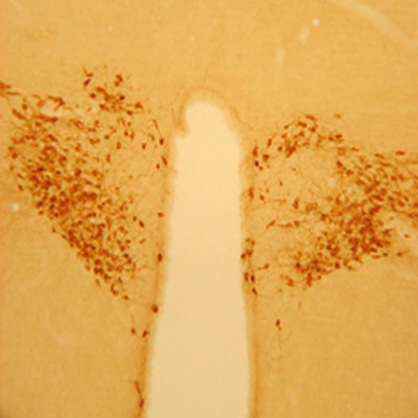
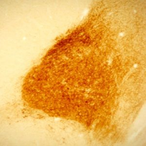
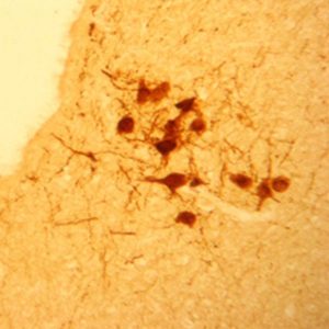
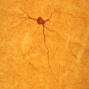
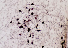
Photo Description: IHC image of neurons staining for oxytocin in the rat hypothalamus (above) and higher magnification of the rat hypothalamus with nickel preparation (below). The tissue was fixed with 4% formaldehyde in 0.1 M phosphate buffer, before being removed and prepared for vibratome sectioning. Floating sections were incubated at RT in 10% goat serum in PBS, before standard IHC procedure. Primary antibody was incubated at 1:5000 for 48 hours, goat anti-rabbit secondary was subsequently added for 1 hour after washing with PBS. Light microscopy staining was achieved with standard biotin-streptavidin/HRP procedure and DAB chromogen.
Host: Rabbit
Quantity / Volume: 100 µL
State: Lyophilized Whole Serum
Reacts With: Bat, Bovine, Chicken, Dog, Frog (Xenopus Laevis), Guinea Pig, Hamster, Human, Manducta Sexta (Moth), Melopsittacus Undulatus (Bird), Mole Rat, Monkey, Mouse, Rat, Sheep, Vole
Availability: In Stock
Alternate Names: Oxytocin-neurophysin 1; Ocytocin; neurophysin I; oxytocin, prepropeptide; oxytocin, prepro- (neurophysin I); oxytocin-neurophysin I, preproprotein; OT; OT-NPI; OXT-NPI; oxytocin/neurophysin I prepropeptide, anti-Oxytocin
Gene Symbol: OXT
RRID: AB_572258
Database Links:
Entrez Gene: 280888 Bovine
Entrez Gene: 768516 Chicken
Entrez Gene: 100856247 Dog
Entrez Gene: 100485685 Frog (Xenopus Laevis)
Entrez Gene: 100720178 Guinea Pig
Entrez Gene: 5020 Human
Entrez Gene: 101717385 Mole Rat
Entrez Gene: 717068 Monkey
Entrez Gene: 18429 Mouse
Entrez Gene: 25504 Rat
Entrez Gene: 443390 Sheep
Entrez Gene: 101993187 Vole
Technical Sheets
This product contains the preservative sodium azide. The concentration percent of the sodium azide is ≤ .09%. Although this hazardous substance is a concentration below that required for the preparation of a Material Safety Data Sheet, we created a standard MSDS for your records.
Download Data SheetDownload MSDS
Reviews
Want to leave a review? Please click here to send us your review.
Beautiful staining
From Robin Forbes-Lormanthe at Ripon College
Beautiful staining in 4% paraformaldehyde fixed rat brain tissue with vector ABC/SG) using a passive perfusion technique and then sectioned coronally with 40 µm thickness.
Works in Prairie Vole Tissues
From the Grippo Lab at Northern Illinois University
We used this oxytocin antibody on prairie vole (Microtus ochrogaster) tissues. Tissues were fixed with 4% paraformaldehyde (with 10% acrolein) using a passive perfusion technique and then sectioned coronally with 40 µm thickness.
We followed an avidin-biotin complex method that was previously published by our laboratory (Grippo, Gerena, et al., 2007). The primary antibody for oxytocin was incubated at the concentration of 1:100,000 at 4ºC for ~70 hours.
The staining was not visible to the naked eye (compared to our previous oxytocin antibody); however, stained cells were visible under the microscope. We did observe some variability of staining within the same dish, but cells were still countable.
Additionally, we also used this antibody in our cFos + oxytocin co-labeling protocol, yielding similar results. These findings will be presented at the 2019 Society for Neuroscience conference (Watanasriyakul et al., 2019).
Overall, it’s a good antibody that works well even at a low concentration.
Excellent for IF
Excellent signal in mouse hypothalamus when used at 1:2000. Brains were first perfused with PBS and then with 4% PFA followed by overnight drop-fixation in 4% PFA. 30um-thick slices were cut on a sliding blade microtome and then immunostained as floating sections. Slices were incubated in the primary anti-oxytocin antibody overnight rocking at 4oC. Primary antibodies were visualized with anti-rabbit secondaries, incubation for 1h at RT the following day.
Would recommend.
This antibody worked very well at 1:2,000 in PFA-fixed, cryosectioned mouse brain tissue. OXT-positive cells in the PVN were very clearly labeled with minimal background.
Not useful for experiments in nonmammalian species
As stated on the antibody product page, this antibody exhibits reactivity to forms of oxytocin across a wide range of species, including species that express Ile8-oxytocin (mesotocin), the oxytocin form found in most marsupials, birds, reptiles and amphibians.
Published work in birds (Cantwell and Cassone; PubMed ID: 1699890; cited on the antibody product page) demonstrates that preadsorption with mesotocin does in fact eliminate immunolabeling in birds. However, those authors did not conduct preadsorptions with the closely related neuropeptide vasotocin, which differs from mesotocin by only one amino acid. Vasotocin is the nonmammalian homologue of vasopressin and is the ancestral peptide for all vertebrate peptides of the oxytocin-vasopressin family. Importantly, vasotocin and mesotocin are expressed in separate subpopulations of neurons in the hypothalamus (e.g., separate subpopulations in the paraventricular nucleus), as are vasopressin and oxytocin, but the oxytocin antibody 20068 labels both, as we have discovered in pilot experiments using a selective vasotocin antibody alongside the oxytocin 20068. The oxytocin antibody also labels extrahypothalamic neurons in the bed nucleus of the stria terminalis that are known to express only vasotocin, not mesotocin (see Goodson et al., 2012, PNAS, 109 supp. 1, 10685-10692).
Thus, although the oxytocin antibody 20068 does recognize nonmammalian forms of oxytocin, it is not useful for experiments in nonmammalian species due to its cross-reactivity to vasotocin, a peptide found in all nonmammalian vertebrates.
————————————————–
Jim Goodson, Professor
Evolution, Ecology and Behavior
Department of Biology
Indiana University
Oxytocin DAB Staining in Rat
I used the Antibody at 1:4000 in rat.
The cell bodies are really easy to see at low magnification throughout the brain. The fibers are hard to see outside of the PVN at anything less than 40x in oil.
Reviewed by:
Yaminah Gilles
Howard University
Neurodevelopmental Biology Laboratory