Description
The antibody has a proven strong biotin-streptavidin/HRP staining at a 1/2000 – 1/4000 dilution in rat hypothalamus.
Staining is completely eliminated by pretreatment of the diluted antibody with 10 µg/mL of arginine vasopressin. Pre-adsorption with as much as 100 µg/mL of oxytocin had no effect on immunolabeling.
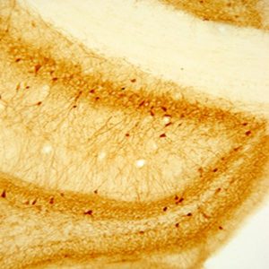
Photo Description: IHC image of rat hypothalamus staining for vasopressin. The tissue was fixed with 4% formaldehyde in phosphate buffer, before being removed and prepared for vibratome sectioning. Floating sections were incubated at RT in 10% goat serum in PBS, before standard IHC procedure. Primary antibody was incubated at 1:5000 for 48 hours, goat anti-rabbit secondary was subsequently added for 1 hour after washing with PBS. Light microscopy staining was achieved with standard biotin-streptavidin/HRP procedure and DAB chromogen.
Host: Rabbit
Quantity / Volume: 100 µL
State: Lyophilized Whole Serum
Reacts With: Anole, Bluehead Wrasses (Fish), Chicken, Clarias Batrachus (Catfish), Cockroach, Ewe / Sheep, Hamster, Human, Hydra Vulgaris (Polyp), Locust, Mole Rat, Monkey, Mouse, Pigeon, Rat, Shrew, Squirrel
Availability: In Stock
Alternate Names: Vasopressin (Arg); Antidiuretic Hormone; Arginine vasopressin; Vasopressin-neurophysin 2-copeptin; ARG-vasopressin; prepro-AVP-NPII; neurohypophyseal; vasopressin-neurophysin II-copeptin; prepro-arginine-vasopressin-neurophysin II; AVP-NPII; VP; ADH; ARVP; AVRP, anti-vasopressin
Gene Symbol: AVP
RRID: AB_572219
Database Links:
Entrez Gene: 396101 Chicken
Entrez Gene: 443477 Ewe / Sheep
Entrez Gene: 551 Human
Entrez Gene: 101717720 Mole Rat
Entrez Gene: 717101 Monkey
Entrez Gene: 11998 Mouse
Entrez Gene: 24221 Rat
Entrez Gene: 102870182 Shrew
Technical Sheets
This product contains the preservative sodium azide. The concentration percent of the sodium azide is ≤ .09%. Although this hazardous substance is a concentration below that required for the preparation of a Material Safety Data Sheet, we created a standard MSDS for your records.
Download Data SheetDownload MSDS
Reviews
Want to leave a review? Please click here to send us your review.
Great antibody
This antibody is the best! I use it for visualizing vasopressin within the rat brain. 1:10,000 dilution results in beautiful staining of the magnocellular regions of the PVN and SON, as well as some staining in the BST and amygdala. My protocol is:
Day 1
_ _ _ Wash 3×5 min TBS
____ 0.1% sodium borohydride in TBS 15 min
_ _ _ Wash 3×5 min TBS
____ 20% normal serum and 3% H202 in TBS 1 hr
____ Primary antibody 1:10K with 2% normal serum in TBS -Triton over 3 nights @ 4º
Send good thoughts over the next 3 days
Day 2
_ _ _ Wash 3×5 min TBS- Triton
____ Secondary antibody 1:500 in TBS-Triton w/ 0.5% gelatin 90 min
____ Make ABC during last 20 min of 2o (25 ul each of A & B per 10 ml TBS)
_ _ _ Wash 1×5 min TBS- Triton, 2×5 min TBS
____ ABC 60 min
_ _ _ Wash 3×5 min TBS-Triton
____ Visualize with Vector SG (6 drops of each reagent per 10 ml TBS-T) + 0.5% gelatin
_ _ _ Wash 3×5 min TBS
Submitted by:
Robin Forbes-Lorman
Biology Department
Ripon College
Ripon, WI
Excellent for IHC
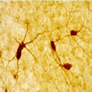
This is a high-quality antibody that we have found to consistently produce beautiful, specific staining. We used a 1:1,000 concentration in cryosectioned mouse brain slices following blocking with 10% serum. Labeled cell bodies and fibers were clearly discernable at 20x magnification in the hypothalamus. Highly recommended.
Beautiful cells
We have used your Vasopressin antibody (Product ID: 20069) in many of our studies and have found in to be a very specific and clear antibody. The staining is great and the cells are beautiful.
We use a total 1:5000 concentration. It is mixed with 10% serum, 10% biotin diluted in 80% PBS-T and incubated for 48-72 hours at 4 degrees C. We used brain tissue from Thamnophis sirtalis (Red-sided garter snake).
Submitted by:
Ashley Maine
Lutterschmidt Lab
Portland State University
Department of Biology
Portland, OR 97207