Description
For induction of c-fos protein activity rats were injected with 1.0 ml of 1.5 M NaCl per 100 grams of body weight. Negative control rats were injected with the same volume of normal saline. The ImmunoStar c-fos antiserum was quality control tested using standard immunohistochemical methods. The antiserum demonstrates strongly positive labeling of rat paraventricular nucleus and supraoptic nucleus using indirect immunofluorescent and biotin/avidin-HRP techniques. No labeling was seen in negative control rats. Recommended primary dilutions for these methods are 1/4000-1/6000 in PBS/0.3% Triton X-100 – FITC Technique and 1/4000-1/6000 in PBS/0.3% Triton X-100 – biotin/avidin-HRP Technique.
Injection of intraperitoneal hypertonic saline to provoke fos expression is most usually used to set ICC staining parameters. It is critical that tissue is harvested 60-90 minutes post injection for this and any other stress that is used. Inject 1 ml/100 g body weight 1.5 M NaCl intraperitoneally. Harvest brains 90 minutes post injection. Time is critical.
Specificity of the antiserum was demonstrated by blockage of staining in experimental rats by omission of c-fos antibody or by substitution of antibody pre-incubated with synthetic peptide or the conjugate. Immunoblot analysis of mediobasal hypothalamus showed a single band of approximately 55-60 kD.
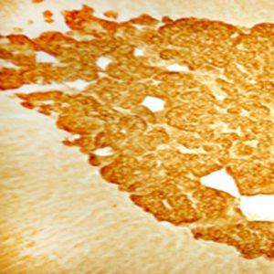
Photo Description: IHC image of neurons staining for c-Fos in the rat supraoptic nucleus. The tissue was fixed with 4% formaldehyde in 0.1 M phosphate buffer, before being removed and prepared for vibratome sectioning. Floating sections were incubated at RT in 10% goat serum in PBS, before standard IHC procedure. Primary antibody was incubated at 1:5000 for 48 hours, goat anti-rabbit secondary was subsequently added for 1 hour after washing with PBS. Light microscopy staining was achieved with standard biotin-streptavidin/HRP procedure and DAB chromogen. The section was then mounted on slides with permount.
Host: Rabbit
Quantity / Volume: 100 µL
State: Lyophilized Whole Serum
Reacts With: Mouse, Rat
Availability: In Stock
Alternate Names: Proto-oncogene c-Fos; Cellular oncogene; G0/G1 switch regulatory protein 7; Proto-oncogene protein; G0S7; p55; AP-1; FBJ murine osteosarcoma viral oncogene homolog, anti-CFOS
Gene Symbol: FOS
RRID: AB_572267
Database Links:
Entrez Gene: 2353 Human
Entrez Gene: 14281 Mouse
Entrez Gene: 314322 Rat
Technical Sheets
This product contains the preservative sodium azide. The concentration percent of the sodium azide is ≤ .09%. Although this hazardous substance is a concentration below that required for the preparation of a Material Safety Data Sheet, we created a standard MSDS for your records.
Download Data SheetDownload MSDS
Reviews
Want to leave a review? Please click here to send us your review.
Beautifully Expressed ir-Fos Cells
We have been using your antibody for years. The consistency and replicability is hands down the best. In fact, whenever we want to try a new antibody, Immunostar is the first company we check for our new antibody of interest.
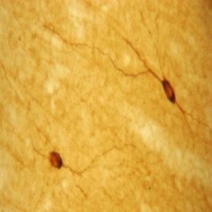
We are using the cFos #26209 antibody on rat brain tissue to look at activity in response to unpredictability. The first step is to perform an antigen retrieval step, placing brains in sodium citrate at 90oC for 15 minutes followed by a peroxidase quench in hydrogen peroxide for 15 minutes and then blocking for an hour before placing in primary antibody overnight at 4oC for 24hrs at a concentrations of 1:10,000. Next the tissue is incubated in secondary antibody for 2hrs at room temperature and then placed in a vectastain ABC solution for 2hrs before the precipitation reaction with DAB and hydrogen peroxide.
The resulting tissue has beautifully expressed ir-Fos cells that are purple because of the added nickel chloride to the DAB. For our purposes we know the staining is specific because we can see staining not only in the hippocampus but also in the amygdala but little specific staining throughout the rest of the brain.
Below are two of our most current staining using the Immunostar antibody.
Kent, M., Scott, S., Kirk, E., Thompson, B., McKearney, N., Dozier, B., Lambert, S., Terhune-Cotter, B., Neal, S., McBardi, M., & Lambert, K. (2018) Contingency training influences neurobiological responses to environmental threats and cognitive uncertainty in male and female rats: A potential rat model for behavioral therapy. Neuroscience: in press.
Kent, M., Bardi, M., Hazelgrove, A., Sewell, K., Kirk, E., Thompson, B., Trexler, K., Terhune-Cotter, B & Lambert, K. (2017) Profiling coping strategies in male and female rats: potential beurobehavioral markers of increased resilience to depressive symptoms. Hormones and Behavior. 95:33-43.
Thanks so much,
Molly Kent
Post-Doctoral Fellow
University of Richmond
Staining Looks Great
My lab recently decided to switch from Santa Cruz to Immunostar for C-fos immunohistochemistry; we tried a Millipore option too with no luck. However, Immunostar came recommended so I ordered Rabbit polyclonal C-Fos antibody, Product ID 26209. I initially tried to adapt our previous protocol and substituted the new primary however after a few tries at various concentrations I was not having any luck. I then contacted Immunostar who was able to put us in contact with another group that had success and was willing to share their protocol, and after a couple modifications, I was able to get the protocol below to work. Overall, the staining looks great and I really appreciate the great customer service.
Fluorescent immunohistochemistry on free-floating sections.
40 micron sections
1.Wash sections in 1X PBS for 3X5 min.
2.Incubate in PBS with 1% Triton-X 100 for 1 hour at RT.
3.Wash in PBS 3X5 min.
4.Incubate for 1 hour at RT in BLOCK.
5.Incubate with primary antibody 3 days in BLOCK. (In fridge)
6.Wash in PBS 3X5 min.
7.Incubate in secondary antibody overnight in BLOCK. (covered in foil in fridge)
8.Wash in PBS 3X5 min. (covered in foil)
9.Mount. Keep slides covered in foil until dry.
10.Coverslip. Use 140ul Vectashield on tissue, and return to foil for storage in fridge.
BLOCK
1X PBS
2% serum of secondary antibody(goat)
0.2% Triton X-100
Primary antibody
Anti-C-Fos (Rabbit): 1:1000
Secondary antibody
Anti-rabbit 568 (red) (Alexa Fluor, abcam) 1:100
Anti-rabbit 488 (green) (Alexa Fluor, abcam) 1:100
Submitted by:
Graduate Student: Rheall Roquet
Principal Investigator: Marie Monfils
University of Texas at Austin
Worked Beautifully
I recently ordered Rabbit polyclonal C-Fos antibody, Product ID 26209 (lot 113018B) from ImmunoStar. The Millipore C-Fos antibody that my laboratory has used for years was no longer available and we had tried 4 other C-fos antibodies with poor results before we found the ImmunoStar C-Fos, which worked beautifully. We found papers in the literature using the ImmunoStar antibody, ABC kit followed by DAB. We used a fluorescent secondary antibody. Protocol is below.
Fluorescent immunohistochemistry on free-floating forebrain and brainstem sections.
Transcardially perfused rats: heparinized (50 U/ml) 0.01 M cold PBS or DMEM (~ 150 ml), then 4% PFA (~ 200 – 250 ml); post-fixed overnight (4% PFA); cryoprotected with 30% sucrose in 0.01M PBS (3 – 5 days); sectioned (35 microns) on a cryostat.
Sections rinsed (0.01M PBS) and blocked in 10% normal donkey serum (NDS) in 0.01M PBS (30 min)
Primary incubation: Overnight on shaker: Room temperature, free-floating sections, 1:3000 dilution of rabbit anti-C-Fos in 0.01 M PBS with 3% NDS and 0.3% Triton.
Secondary: 2 hrs, room temperature, donkey anti-rabbit IgG Alexa-488 (Jackson laboratories). 1:200 dilution in 0.01M PBS with 3% NDS.
Mounted: Gel coated slides; dried; coverslipped using Prolong Gold
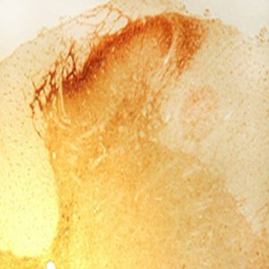
Staining: Trial with hypertonic saline treated rat: Defined round nucei, with evident nucleolus in activated cells in the hypothalamic paraventricular nucleus. Good contrast, no background staining.
Submitted by:
Cheryl Heesch,
University of Missouri,
Columbia, Missouri, 65211