Description
The ImmunoStar serotonin antiserum was quality control tested using standard immunohistochemical methods. The antiserum demonstrates strongly positive labeling of rat hypothalamus and spinal cord using indirect immunofluorescent and biotin/avidin-HRP techniques. Recommended primary dilution is 1/5000 – 1/10,000 in PBS/0.3% Triton X-100 – Bn/Av-HRP Technique.
Staining is completely eliminated by pretreatment of the diluted antibody with 100 µg of serotonin/BSA conjugate.
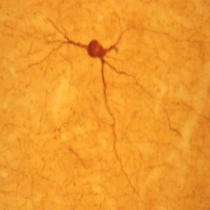
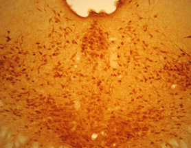
Photo Description: Low magnification of IHC image of neurons staining for the 5-HT goat antibody in the pontine region of the rat brain (top), and high magnification of the raphe nucleus (bottom). The tissue was fixed with 4% formaldehyde/0.05% glutaraldehyde in 0.1 M phosphate buffer, before being removed and prepared for vibratome sectioning. Floating sections were incubated at RT in 10% rabbit serum in PBS, before standard IHC procedure. Primary antibody was incubated at 1:7500 for 48 hours, rabbit anti-goat secondary was subsequently added for 1 hour after washing with PBS. Light microscopy staining was achieved with standard biotin-streptavidin/HRP procedure and DAB chromogen.
Host: Goat
Quantity / Volume: 100 µL
State: Lyophilized Whole Serum
Availability: In Stock
Reacts With: Cat, Cod, Cow, Crayfish, Dolphin, Fish, Frog (Xenopus Laevis), Guinea Pig, Hamster, Human, Mexican salamander (Ambystoma Mexicanum), Monkey, Moth, Mouse, Pig, Rabbit, Ram (Sheep), Rat, Salamander, Snail, Tapeworm, Worm, Zebrafish
Alternate Names: anti-5-HT Goat
RRID: AB_572262
Immunogen: Serotonin
Gene Symbol: Spl
Gene ID: 104153
Technical Sheets
This product contains the preservative sodium azide. The concentration percent of the sodium azide is ≤ .09%. Although this hazardous substance is a concentration below that required for the preparation of a Material Safety Data Sheet, we created a standard MSDS for your records.
Download Data SheetDownload MSDS
Reviews
Want to leave a review? Please click here to send us your review.
Beautiful Axonal Labeling
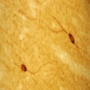
We are using goat 5HT antibody on 20 µm rat spinal cord direct mounted sections. Slides are incubated overnight with anti 5HT at 1:500, rinsed with TBS, followed by donkey-anti goat–Alexafluor 568 at 1:250 for 3 hours.
Very reliable antibody with beautiful axonal labeling.
Submitted by:
Dr. Yacov Koffler
Project Scientist
Center for Neural Repair
UC San Diego
This serotonin antiserum is great
Rat brain tissues were perfused with 4% paraformaldehyde in 0.1 M PBS (PH 7.6). The tissues were equilibrated in cold 25% sucrose, sectioned (10 uM thick) in a microtome and stored at -20 C prior to immunohistochemical assay.
Brain tissue sections were rinsed three times in 0.1 M PBS and then incubated with ImmunoStar 5-HT (Serotonin) Goat Antibody (Item # 20079) diluted 1:5000 overnight at 4C. After rinsing with 0.1 M PBS, the sections were incubated an anti-rabbit secondary antiserum for 2 h. This secondary antiserum has been shown to have minimal cross-reactivity to other relevant species. Sections were cover slipped and examined under a confocal microscope system.
Brain tissue sections had the clear 5-HT immunolabeling in serotonin neuron dendrites while the negative control sections (without 5-HT (Serotonin) Goat Antibody treatment) showed just background fluorescence. This serotonin antiserum is great and it did work beautifully in our set-up.
Jason Zhang
Project Scientist
University of California-San Diego
Very Good Antibody – Recommended
We use ImmunoStar 5-HT goat antibody (ID #20079) to stain brain tissue of a marine fish. We combine it with fluorescent-labeled secondary antibodies for visualization under epifluorescence microscopy.
This antibody produces a robust signal with minimal background and clear staining of somata and processes. Our neuroanatomy results with ImmunoStar goat anti-serotonin are identical to those we found using another well known antibody from another manufacturer made in rabbit.
Brain tissue is from animals transcardially perfused with Ringer’s solution followed by 4% paraformaldehyde in phosphate buffer (0.1M, pH 7.2). Tissue is cryosectioned at 25 microns.
Our procedure is as follows:
1. Rehydrate tissue in PBS (0.1M phosphate buffer saline, pH 7.2) for 10 minutes twice.
2. Immerse tissue in PBST+DS-5% (PBS with 5% donkey serum and 0.3% Triton-X) for 1 hour.
3. Apply goat 5-HT antiserum diluted 1:1000 in PBST+DS-5% and incubate at room temperature overnight.
4. Wash tissue with PBS+DS-0.5% (0.5% donkey serum solution, no Triton-X) for 10 minutes five times.
5. Apply secondary antiserum diluted 1:400 in PBST+DS-5% and incubate at room temperature for 2 hours.
6. Wash with PBS for 10 minutes six times.
7. Mount slides and view.
Miky Timothy
Graduate Research Assistant
Department of Biology
Brooklyn College – The City University of New York
Very Reliable Antibody
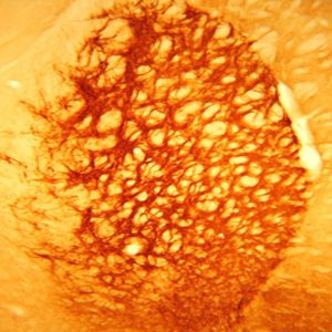
I find ImmunoStar’s goat anti-serotonin (#20079) to be just as reliable as the rabbit anti-serotonin antibody. I’ve used this on sectioned brain tissue from moths, flies, cockroaches and preying mantids. I always get very even penetration of the antibody although I have never tried thicker than 150um sections of brain embedded in agarose blocks.
Below is my protocol. I also have a picture of the brain of a cockroach labeled with this antibody.
Day 1
Overnight (O/N) at 4oC in 4% paraformaldehyde in 0.01 Phosphate Buffered Saline (PBS).
Day 2
Wash tissue 3x 5 min in PBS. Embed in 5% agarose and section at 50-150um.
Wash 4x 15 min in PBS with .5% Triton X-100 (PBST). Block 1 hr in PBST with 2% Bovine Serum Albumin (BSA). O/N at room temperature (RT) in 1:1000 goat anti-5HT in PBST with 5mM sodium azide (PBSAT) with 2% BSA and 1% Triton X-100.
Day 3
Wash 4x 30 min PBST. Block 1hr 2% BSA in PBST. O/N at RT in 1:1000 donkey anti-goat Alexa 546 (2% BSA in PBSAT).
Day 4
Wash 2x 15min PBST. Wash 2x 15min PBS. Wash 1x 15min in 60% glycerol. Mount in 80% glycerol.
Andrew Dacks, Ph.D.
Assistant Professor
Department of Biology
West Virginia University
Great staining with Serotonin antibody
I utilized the antibody in formalin-fixed, free-floating mouse brain tissue at a concentration of 1:1000. For secondary detection I utilized Invitrogen Alexa-Fluor-conjugated secondary antibodies at 1:200.
The 5HT antibody always yielded great staining and we were very pleased with it. Indeed, we utilized this antibody for studies of dorsal raphe 5HT neurons as reported in our recent publication in Cell Metabolism (Lam, Leinnintger et al 2011, Cell Metab 13(5): 584-91).
Gina M. Leinninger, PhD
Research Investigator
Martin G. Myers Jr. Laboratory
University of Michigan