Description
The antibody is provided as 100 uL of affinity purified serum in PBS (0.02 M sodium phosphate with 0.15 M sodium chloride, pH 7.5) with 1% BSA (bovine serum albumin), and 0.02% sodium azide. Reacts with mouse, rat, and rabbit species.
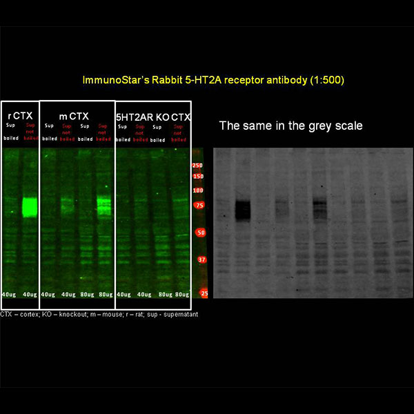
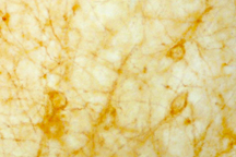
The images above labeled “ImmunoStar Rabbit 5-HT 2A receptor antibody” are the results of staining of the mouse 5-HT 2A receptor in the brain, courtesy of Dr. Magdalena Zaniewska, Max-Delbruck-Centrum fur Molekulare Medizin Berlin, Germany.
The ImmunoStar 5HT 2A receptor antibody was quality control tested using standard immunohistochemical methods. The antiserum demonstrates strongly positive labeling of rat cortex, amygdala and hippocampus using indirect immunofluorescent and biotin/avidin-HRP techniques. Recommended primary dilutions are 1/300 – 1/500 in PBS/0.3% Triton X-100 – Bn/Av-HRP Technique . The addition of intensifying reagents such as nickel ammonium sulfate to the chromogen solution will approximately double the dilution factor as recommended.
Immunolabeling is completely abolished by preadsorption with synthetic rat 5HT2A receptor (22-41). Immunolabeling of Western blot revealed a single band of approximately 53kD. Due to the difficulty with receptor antibodies, western blot applications are not warranted and are included as specificity information only.
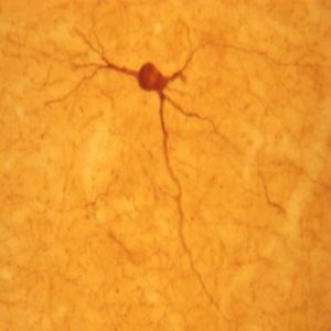
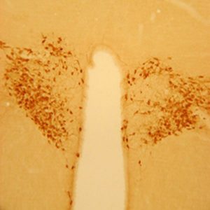
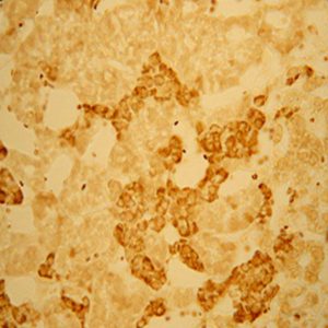
Photo Description: Low magnification IHC image of neurons staining for the 5-HT2A receptor in the rat cortex (top of page) and image of neuronal expression of the receptor in the amygdala (below). The bottom right photo is of the cortex. The tissue was fixed with 4% formaldehyde in 0.1 M phosphate buffer, before being removed and prepared for vibratome sectioning. Floating sections were incubated at RT in 10% goat serum in PBS, before standard IHC procedure. Primary antibody was incubated at 1:500 for 48 hours, goat anti-rabbit secondary was subsequently added for 1 hour after washing with PBS. Light microscopy staining was achieved with standard biotin-streptavidin/HRP procedure and DAB chromogen.

Host: Rabbit
Quantity / Volume: 100 uL
State: Liquid
Reacts With: Human, Mouse, Rabbit, Rat
Alternate Names: 5-hydroxytryptamine receptor 2A; 5Ht-2; 5-hydroxytryptamine (serotonin) receptor 2A, G protein-coupled, anti-5-HT 2A
Gene Symbol Htr2a
Database Links:
Entrez Gene: 3356 Human
Entrez Gene: 15558 Mouse
Entrez Gene: 100009461 Rabbit
Entrez Gene: 29595 Rat
RRID: AB_572211
Technical Sheets
This product contains the preservative sodium azide. The concentration percent of the sodium azide is ≤ .09%. Although this hazardous substance is a concentration below that required for the preparation of a Material Safety Data Sheet, we created a standard MSDS for your records.
Download Data SheetDownload MSDS
Reviews
Want to leave a review? Please click here to send us your review.
Only your AB shows a specific signal
(Please refer to the above western blot image submitted by Dr. Schwendt.)
We have been interested in detecting changes of 5-HT2a in rodent brain, primarily by immunoblotting, also by immunohistochemistry. We have tested several commercial antibodies, with only your Ab showing a specific signal, as verified by brain distribution, preadsorbtion and by a lack of signal in KO animals.
We have optimized a immunoblotting protocol which works well to detect 5-HT2a in rat brain lysates. Mouse tissue is a little bit more difficult but still possible
Immunoblotting: Rat or mouse brain samples were lysed by brief sonication in a buffer containing: 1% SDS in 1xPBS, protease inhibitors (Complete Mini, Roche). Samples were mixed with a modified Leamli buffer (containing a reducing agent; 40mM DTT), incubated at 37C for 30mins and separated by SDS-PAGE. Separated proteins were then blotted onto a PVDF membrane, blocked with 5% milk/TBS/0.1% Tween80 and probed with anti-5HT2a antibody (1:500, Immunostar #24288) followed by anti-rabbit HRP- conjugated secondary antibody (1:5000, Jackson Immuno). Signal was visualized using ECL2 kit according to manufacturer’s instructions (Thermo).
Image Samples: 1 (Rat prefrontal cortex, 25ug), 2 (Rat laterodorsal thalamus, 25ug), 3 (Wt mouse cortex, 25 ug), 4 (5-HT2a KO mouse cortex, 25 ug), 5 (Wt mouse cortex, 75 ug), 6 (5-HT2a KO mouse cortex, 75 ug). As expected, sample in lane 2 showed low expression, and samples in lanes 4 and 6 showed no expression of 5-HT2a receptors. Mouse tissues obtained from Dr. Gonzalez-Maeso (Mount Sinai Hospital, NY) indicates detected 5Ht2a receptor signal.
Submitted by:
Marek Schwendt, PhD
Psychology Department
University of Florida
Gainesville, Fl
Antibodies are specific for mouse receptor
[The two pictures above labeled “ImmunoStar’s Rabbit 5-HT 2A receptor antibody” are courtesy of Dr. Magdalena Zaniewska.]
Here are the results of staining of the mouse 5-HT 2A receptors in the brain with the use of Immunostar’s antibodies (Cat no: 24288). I think the antibodies are specific for the mouse receptor (no receptor labeling in the brain of 5-HT 2A receptor knockout mouse) and can be used in a Western blot. What I already found out is that 5-HT 2A receptors in the cortex seem to be glycosylated (band around 75 kDa) since incubation with PNGaseF releases the protein that is around 50 kDa.
We received the 5-HT 2A receptor knockout animals from Dr. Joelle Adrien from another research team in Paris (see below).
Reviewed by:
Dr. Magdalena Zaniewska
Molekularbiologie von Hormonen im Herz-Kreislaufssystem
Max-Delbrϋck-Centrum fϋr Molekulare Medizin
Berlin, Germany
Dr. Joelle Adrien
Directeur de Recherches a l’Inserm
Faculte de Medecine Pierre et Marie Curie – Site Pitie Salpetriere
Paris, France