Description
The ImmunoStar Neurotensin antiserum was quality control tested using standard immunohistochemical methods. The antiserum demonstrates strongly positive labeling of rat amygdala using indirect immunofluorescent and biotin/avidin-HRP techniques. Recommended primary dilution is 1/4000-1/8000 in PBS/0.3% Triton x-100 – Bn/Av-HRP Technique. Staining is completely eliminated by pretreatment with 10 µg of Neurotensin per 1 mL of diluted antibody.
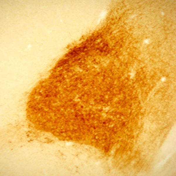
Photo Description: IHC image of rat amygdala staining for neurotensin. The tissue was fixed with 4% formaldehyde in 0.1 M phosphate buffer, before being removed and prepared for vibratome sectioning. Floating sections were incubated at RT in 10% goat serum in PBS, before standard IHC procedure. Primary antibody was incubated at 1:5000 for 48 hours, goat anti-rabbit secondary was subsequently added for 1 hour after washing with PBS. Light microscopy staining was achieved with standard biotin-streptavidin/HRP procedure and DAB chromogen.
Host: Rabbit
Quantity / Volume: 100 µL
State: Lyophilized Whole Serum
Reacts With: Cat, Dog, Frog, Human, Monkey, Mouse, Pigeon, Rat, Turtle
Availability: In Stock
Alternate Names: neuromedin N; Preproneurotensin; pro-neurotensin, anti-neurotensin
Gene Symbol: NTS
RRID: AB_572254
Database Links:
Entrez Gene: 101095835 Cat
Entrez Gene: 611687 Dog
Entrez Gene: 100485937 Frog
Entrez Gene: 4922 Human
Entrez Gene: 700189 Monkey
Entrez Gene: 67405 Mouse
Entrez Gene: 102087679 Pigeon
Entrez Gene: 299757 Rat
Entrez Gene:102446489 Turtle
Technical Sheets
This product contains the preservative sodium azide. The concentration percent of the sodium azide is ≤ .09%. Although this hazardous substance is a concentration below that required for the preparation of a Material Safety Data Sheet, we created a standard MSDS for your records.
Download Data SheetDownload MSDS
Reviews
Want to leave a review? Please click here to send us your review.
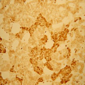
Staining for Ventromedial Ventral Pallidum
We used the rabbit anti-neurotensin antibody for staining of ventromedial ventral pallidum in rats who were observed during cocaine self administration.
Rats were perfused with saline followed by 4% PFA and stored in 20% sucrose-azide. Brains were coronally sectioned to 40 microns and stored in 0.1 M PBS-Azide (pH 7.4).
The following protocol for IHC staining was used: Free floating sections of rat brain were blocked in 1% hydrogen peroxide in 0.1M PBS for 15 minutes. After washing, tissue was blocked in 0.1M PBS-Triton X-100 (PBST) and 2% normal donkey serum (NDS) for 2 hours. After washing, primary antibody was used in a range of concentrations (1:2500, 1:5000, 1:10000, 1:20000) overnight in PBST and 2% NDS. After washing, secondary antibody used was biotinylated donkey anti-rabbit (Jackson ImmunoResearch) in a 1:500 dilution in PBST over 2 hours. After wash, tissue placed in ABC peroxide (kit from Vecta Stain) 1:500 in PBST for 1.5 hours. Tissue was developed in brown DAB for 10 minutes.
The borders of the ventromedial VP were very clear with this antibody and with low background. Even with concentrations ranging from 1:2500 to 1:20,000, the subregion borders were very easy to identify without having to perform the DAB reaction for an extended period of time.
Christina Ruiz
Lab Manager, SV Mahler Lab
UC Irvine, Department of Neurobiology and Behavior
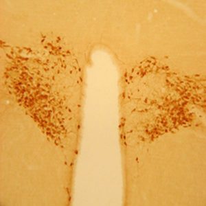
Visualization of ventromedial ventral pallidum
For our manuscript, Differential roles of ventral pallidum subregions during cocaine self-administration behaviors, in press at Journal of Comparative Neurology, we used the Immunostar rabbit anti-neurotensin primary antibody to visualize the ventromedial ventral pallidum subregion.
Rats were perfused with saline followed by 4% PF, stored in 30% sucrose, and coronally sectioned to 40 um. We followed the protocols of Zahm/Heimer and colleagues when they discovered the ventral pallidum subregions and their afferent/efferent projection patterns.
The protocol consisted of the following steps:
1. Wash in 0.1M phosphate buffer (PB; pH 7.4)
2. 15 min in 1% sodium borohydride
3. Wash
4. 1 hour blocking with PB containing 0.1% Triton X-100 and 3% normal goat serum
5. Primary antibody overnight at 4C – ImmunoStar rabbit anti-neurotensin diluted 1 : 6500 in PB containing 0.1% Triton X-100 and 3% normal goal serum
6. Wash
7. 1 hour anti-rabbit biotinylated secondary (Vector) 1:200 in PB with 0.1% Triton X-100
8. Wash
9. 1 hour ABC (Vector) 1:200 in PB with 0.1% Triton X-100
10. Wash
11. Develop with 0.05% DAB for 6 min
Our study involved recording neurons within the ventral pallidum subregions during specific aspects of intravenous cocaine self-administration (approaching toward, responding on, or retreating away from a cocaine-reinforced operandum). In order to verify the placement of microwires within the VP subregions, prior to perfusion we passed current through each stainless steel microwire to leave an iron deposit at the uninsulated tip. After DAB (brown reaction) and mounting, we visualized the iron deposit by incubating in a 5% potassium ferrocyanide and 10% HCl solution (leaving a blue-green reaction).
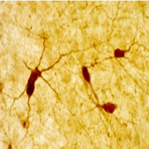
For our purposes, this antibody labeled fibers in ventromedial VP with low background. Individual neurons and other fibers were clearly observed within BNST that were not involved in our study.
I rated this antibody 4 stars because there was some variability for this stain between animals.
David H. Root, Ph.D.
Rutgers University
Crisp, Cell-specific labeling
Tissue: Maccaca fasciscularis, perfused with saline and 4% paraformaldehyde, dehydrated through increase sucrose gradients, sectioned at 40 um with sliding microtome.
Method: Immunocytochemistry on free floating sections. Incubated with ImmunoStar’s primary antibody at a concentration of 1:1000 for 4 days in 10% normal goat serum (in 0.1M PO4 buffer with .3% Triton X-100). Rinsed, then incubated with Vector Lab’s biotinylated goat anti-rabbit secondary antibody (#BA-1000) at a concentration of 1:200 for 40 minutes at room temperature in the same 10% normal goat serum solution. Rinsed, then incubated with Vector’s standard peroxidase kit (#PK-4000) for 60 minutes at room temperature in 0.1M PO4 buffer with .3% Triton X-100. Rinsed, then developed using the instructions for the kit.
Results: Crisp, cell-specific labeling. Low background. Very helpful for determining the boundary between the medial and lateral core divisions of the central nucleus of the amygdala.
Daniel Tylee
Department of Neurobiology & Anatomy
University of Rochester – School of Medicine and Dentistry