Description
Neuropeptide Y (NPY) is a member of a regulatory peptide family and has a marked sequence homology with pancreatic polypeptide (PP) and peptide YY (PYY), which are other members of the family. NPY is widely expressed in the nervous system and has been shown to be differentially expressed in inhibitory interneurons in the hippocampus in degenerative disease, as a powerful vasoconstrictor in the periphery, and increased expression of NPY in the hypothalamus correlates with food intake.
In the rat central nervous system, immunohistochemistry has found NPY-like cell bodies in the cortex, caudate-putamen, hypothalamus (arcuate nucleus), hippocampus, anterior westolfactory bulb, nucleus accumbens, amygdaloid complex and periaqueductal grey. NPY-like fibers and terminals are detected in high numbers in the bed nucleus of the stria terminalis, the peri- and paraventricular regions of the hypothalamus and thalamus and in discrete hypothalamic nuclei, particularly the suprachiasmatic nucleus.
Westerns are not recommended for the Neuropeptide Y antibody since the protein is too small to detect in gel.
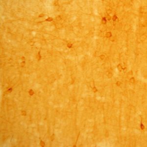
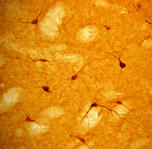
Photo Description: IHC image of neurons staining for neuropeptide Y in the rat cortex (above) and striatum (below). The tissue was fixed with 4% formaldehyde in phosphate buffer, before being removed and prepared for vibratome sectioning. Floating sections were incubated at RT in 10% goat serum in PBS, before standard IHC procedure. Primary antibody was incubated at 1:5000 for 48 hours, goat anti-rabbit secondary was subsequently added for 1 hour after washing with PBS. Light microscopy staining was achieved with standard biotin-streptavidin/HRP procedure and DAB chromogen.
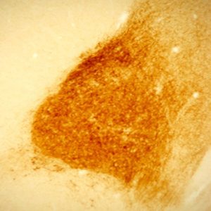
The second image above entitled “C.” is NPY interneurons (RED) in mouse striatum, GREEN staining is GFP expressed by medium spiny neurons provided Alipi Naydenov, Neurobiology and Behavior, Stella Lab University of Washington (see review below).
Host: Rabbit
Quantity / Volume: 100 µL
State: Lyophilized Whole Serum
Reacts With: Alpaca (Llama), Bird, Bubalus Bubalis (Water Buffalo), Bullfrog, Cat, Ewe, Ferret, Fish, Frog, Frog (Xenopus Laevis), Ground Squirrel, Guinea Pig, Hamster, Human, Monkey, Mouse, Ovine (Sheep), Pig, Psetta Maxima (Flat Fish), Rat, Sheep, Snake, Tadpole, Toad, Turtle, Zebrafish
Availability: In Stock
Preabsorption Control: Available at Sigma Aldrich, Catalog # N3266
Alternate Names: Pro-neuropeptide Y; Neuropeptide tyrosine; prepro-neuropeptide Y; PYY4, anti-NPY
Gene Symbol: NPY
RRID: AB_572253
Database Links:
Entrez Gene: 102414548 Bubalus Bubalis (Water Buffalo)
Entrez Gene: 101096968 Cat
Entrez Gene: 443508 Ewe
Entrez Gene: 101686990 Ferret
Entrez Gene: 779985 Frog
Entrez Gene: 101976112 Ground Squirrel
Entrez Gene: 100379621 Guinea Pig
Entrez Gene: 100758436 Hamster
Entrez Gene: 4852 Human
Entrez Gene: 574114 Monkey
Entrez Gene: 109648 Mouse
Entrez Gene: 397304 Pig
Entrez Gene: 24604 Rat
Entrez Gene: 102454441 Turtle
Entrez Gene: 30281 Zebrafish
Technical Sheets
This product contains the preservative sodium azide. The concentration percent of the sodium azide is ≤ .09%. Although this hazardous substance is a concentration below that required for the preparation of a Material Safety Data Sheet, we created a standard MSDS for your records.
Download Data SheetDownload MSDS
Reviews
Want to leave a review? Please click here to send us your review.
Extremely Specific with Little Background Signal
The NPY #22940 is an antibody we use frequently in our resilience phenotyping and effort based reward training studies to show how some coping strategies can have an innate resilient phenotype and how others can be trained to become more resilient.
NPY has been implicated as a peptide for resilience. For all of our research we look to Immunostar for their antibodies and find that the quality control and customer service is unmatched. The suggested concentration for NPY does actually work best and does not bleed the bank dry.
The staining we have been able to get with our Immunostar NPY antibodies has been extremely specific with little background signal. Allowing us to easily quantify the peptide in dendrites, cells bodies, and other locations within brain tissue.
We use the antibody in paraformaldehyde fixed brains. After quenching with a low concentration of hydrogen peroxide for 15 minutes and blocking with normal goat serum for 1 hour, we use a dilution of 1:5,000, incubating for 48 hours at 4oC. Secondary incubation and ABC amplification occur at room temperature for approximately 90 minutes each followed by DAB and nickel chloride color precipitation. Our tissue staining results in small purple fibers in very specific areas. There is a very low signal to noise ratio allowing for an easy degree of quantification.
Below is our most current article using the immunostar NPY antibody.
Kent, M., Bardi, M., Hazelgrove, A., *Sewell, K., *Kirk, E., *Thompson, B., *Trexler, K., *Terhune-Cotter, B & Lambert, K. (2017) Profiling coping strategies in male and female rats: potential neurobehavioral markers of increased resilience to depressive symptoms. Hormones and Behavior. 95:33-43.
Molly Kent
Post-Doctoral Fellow
University of Richmond
Love the results
We have used your NPY(Product ID: 22940) antibody and love the results we obtain with this antibody. While it is not as clear as your Vasopressin antibody (Product ID: 20069), it works very well for our research purposes. For NPY we use a dilution of 1:3000 and amplify with tyramide solution at a 1:50 dilution. It is mixed with 10% serum, 10% biotin diluted in 80% PBS-T and incubated for 48-72 hours at 4 degrees C. We used brain tissue from Thamnophis sirtalis (Red-sided garter snake).
Submitted by:
Ashley Maine
Lutterschmidt Lab
Portland State University
Department of Biology
Portland, OR 97207
Antibody Performed Very Well
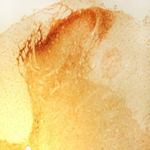
Above IFC image entitled “C.” is NPY interneurons (RED) in mouse striatum, GREEN staining is GFP expressed by medium spiny neurons. NPY antibody was used at 1:1000 dilution with the staining protocol described below. Antibody performed very well. Scale bar = 25 microns.
Immunohistochemistry: Mice were euthanized and perfused with PFA (4% in PBS), post fixed for 24 hrs, and their brains cryoprotected in 15% sucrose (24 hrs) followed by 30% sucrose (48 hrs). Coronal sections that included the corticostriatal, globus pallidus, or substantia nigra regions (30 um) were prepared using a microtome and then stored in PBS at 4C until processing. Sections were processed/stained in parallel as follows: Free floating sections were rinsed 3x with PBS and incubated 90 min at room temperature (RT) in PBS supplemented with donkey serum (5%) and Triton X-100 (1%). Primary antibody combinations were prepared as a master stock in PBS supplemented with donkey serum (2.5%) and Triton X-100 (0.5%) and applied to sections for 72 hrs at 4°C with gentle agitation. Sections were then rinsed 8x with PBS supplemented with Tween-20 (0.05%, at RT) and incubated with a secondary antibodies diluted in PBS supplemented with donkey serum (2.5%) and Triton X-100 (0.5%) for 1 hr at RT with gentle agitation, followed by 7 rinses with PBS and one rinse with deionized water. Sections were mounted onto slides and allowed to dry for ~18 hrs, after which cover slips were mounted with Vectashield and sealed with nail polish.
Review submitted by:
Alipi Naydenov
Neurobiology and Behavior, Stella Lab
University of Washington